Healthy Inspirations

Digital Radiology Technology
Wed, 28 Aug 2024In the medical field, radiology examinations are very helpful for doctors to view the internal parts of a patient’s body in order to gain insights into their medical condition. Nearly every healthcare facility has integrated radiology services. This aims to facilitate coordination between doctors and the radiology department so that patients can be diagnosed and treated effectively.
The radiology examination process uses radiographic equipment with various machines and other techniques to produce images of the structures and activities within the body. The type of imaging used by doctors depends on the symptoms and the part of the body being examined.
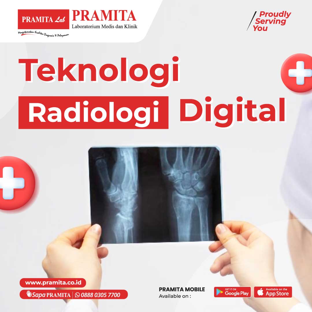
The latest system in radiology examinations includes Direct Digital Radiography (DDR). Digital Radiology uses several key technologies to produce accurate and high-quality radiographic images. The principle of DDR is to capture X-rays without film and replace it with digital devices. With this system, the images from exposure can be viewed on a monitor in real-time, and files can be saved or sent to all devices. Although still relatively new, some healthcare facilities have already adopted this technology for medical diagnosis.
Current DDR (Direct Digital Radiography) equipment consists of main components including an X-ray generator and a detector composed of a fluorescent screen and a digital camera. The detector used in Digital Radiology is capable of converting radiation energy into electronic signals, which are then transformed into digital images. The use of digital systems and computers allows operators or radiographers to apply distance protocols with patients. The system’s performance has also been optimized to produce high-quality digital images.
Generally, the radiographic procedure using DDR is almost the same as conventional film-based radiographic equipment. The fundamental difference lies in the operation and the final process of producing the images from exposure.
Before performing a radiography procedure, the radiographer first ensures that reference images have been obtained, checks the exposure request, and inputs patient data. The radiographer then guides the radiography process and verifies the patient data that has been entered.
For patients, radiography is generally performed in a standing position for chest imaging requests. For other radiography requests, such as plain abdominal X-rays, the patient will be directed to lie on the provided bed or usually a bed for radiography.
The radiographer then sets the exposure parameters for the patient according to the examination to be performed. After the exposure process is complete, the radiographer obtains digital images that can be directly accessed for verification and validation from the server or computer. This equipment also connects a network between healthcare facilities so that patient services can be conducted quickly, efficiently, effectively, and productively.
Author: Dr. Dewi Maria, Sp.Rad (Head of Radiology at PRAMITA Lab Wahid Hasyim branch)

